Insulin
From Wikipedia, the free encyclopedia


Insulin is a hormone with extensive effects on both metabolism and several other body systems (eg, vascular compliance). Insulin causes most of the body's cells to take up glucose from the blood (including liver, muscle, and fat tissue cells), storing it as glycogen in the liver and muscle, and stops use of fat as an energy source. When insulin is absent (or low), glucose is not taken up by most body cells and the body begins to use fat as an energy source (ie, transfer of lipids from adipose tissue to the liver for mobilization as an energy source). As its level is a central metabolic control mechanism, its status is also used as a control signal to other body systems (such as amino acid uptake by body cells). It has several other anabolic effects throughout the body. When control of insulin levels fails, diabetes mellitus results.
Insulin is used medically to treat some forms of diabetes mellitus. Patients with Type 1 diabetes mellitus depend on external insulin (most commonly injected subcutaneously) for their survival because the hormone is no longer produced internally. Patients with Type 2 diabetes mellitus are insulin resistant, and because of that, may suffer from a relative insulin deficiency; some patients with Type 2 diabetes may eventually require insulin when other medications fail to control blood glucose levels adequately.
Insulin is a peptide hormone composed of 51 amino acid residues and has a molecular weight of 5808 Da. It is produced in the islets of Langerhans in the pancreas. The name comes from the Latin insula for "island".
Insulin's structure varies slightly between species of animal. Insulin from animal sources differs somewhat in 'strength' (i.e., in carbohydrate metabolism control effects) in humans because of those variations. Porcine (pig) insulin is especially close to the human version.
Contents |
[edit] Gene
The proinsulin precursor of insulin is encoded by the INS gene.[2][3]
[edit] Alleles
A variety of mutant alleles with changes in the coding region have been identified. There is a read-through gene, INS-IGF2, which overlaps with this gene at the 5' region and with the IGF2 gene at the 3' region.[2]
[edit] Regulation
There are several regulatory sequences in the promoter region of the human insulin gene, to which transcription factors bind.
In general, the A-boxes bind to Pdx1 factors, E-boxes bind to NeuroD, C-boxes bind to MafA and cAMP response elements to CREB.
There are also silencers that inhibit transcription.
| Regulatory sequence | binding transcription factors |
|---|---|
| ILPR | Par1 |
| A5 | Pdx1 |
| negative regulatory element (NRE)[5] | glucocorticoid receptor, Oct1 |
| Z (overlapping NRE and C2) | ISF |
| C2 | Pax4, MafA(?) |
| E2 | USF1/USF2 |
| A3 | Pdx1 |
| CREB RE | - |
| CREB RE | CREB, CREM |
| A2 | - |
| CAAT enhancer binding (CEB) (partly overlapping A2 and C1) | - |
| C1 | - |
| E1 | E2A, NeuroD1, HEB |
| A1 | Pdx1 |
| G1 | - |
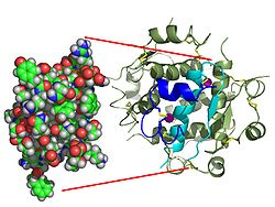
[edit] Protein structure
Within vertebrates, the similarity of insulins is extremely close. Bovine insulin differs from human in only three amino acid residues, and porcine insulin in one. Even insulin from some species of fish is similar enough to human to be clinically effective in humans. Insulin in some invertebrates (eg, the Caenorhabditis elegans nematode) is quite close to human insulin, has similar effects inside cells, and is produced very similarly. Insulin has been strongly preserved over evolutionary time, suggesting its centrality in animal metabolic control. The C-peptide of proinsulin (discussed later), however, differs much more amongst species; it is also a hormone, but a secondary one.
[edit] Synthesis, physiological effects, and degradation
[edit] Synthesis
Insulin is produced in the pancreas, and released when any of several stimuli are detected. These include protein ingestion, and glucose in the blood (from food which produces glucose when digested -- characteristically this is carbohydrate, though not all types produce glucose and so an increase in blood glucose levels). In target cells, they initiate a signal transduction which has the effect of increasing glucose uptake and storage. Finally, insulin is degraded, terminating the response.

In mammals, insulin is synthesized in the pancreas within the beta cells (β-cells) of the islets of Langerhans. One million to three million islets of Langerhans (pancreatic islets) form the endocrine part of the pancreas, which is primarily an exocrine gland. The endocrine portion only accounts for 2% of the total mass of the pancreas. Within the islets of Langerhans, beta cells constitute 60–80% of all the cells.
In beta cells, insulin is synthesized from the proinsulin precursor molecule by the action of proteolytic enzymes, known as prohormone convertases (PC1 and PC2), as well as the exoprotease carboxypeptidase E.[6] These modifications of proinsulin remove the center portion of the molecule (ie, C-peptide), from the C- and N- terminal ends of proinsulin. The remaining polypeptides (51 amino acids in total), the B- and A- chains, are bound together by disulfide bonds/disulphide bonds. Confusingly, the primary sequence of proinsulin goes in the order "B-C-A", since B and A chains were identified on the basis of mass, and the C peptide was discovered after the others.
The endogenous production of insulin is regulated in several steps along the synthesis pathway:
- At transcription from the insulin gene
- In mRNA stability
- At the mRNA translation
- In the posttranslational modifications
It has been shown that insulin and its related proteins, are also produced inside the brain and that reduced levels of these proteins are linked to Alzheimer's disease.[7][8][9]
[edit] Release
Beta cells in the islets of Langerhans release insulin mostly in response to increased blood glucose levels through the following mechanism (see figure to the right):
- Glucose enters the beta cells through the glucose transporter GLUT2
- Glucose goes into the glycolysis and the respiratory cycle where multiple high-energy ATP molecules are produced by oxidation
- Dependent on ATP levels, and hence blood glucose levels, the ATP-controlled potassium channels (K+) close and the cell membrane depolarizes
- On depolarization, voltage controlled calcium channels (Ca2+) open and calcium flows into the cells
- An increased calcium level causes activation of phospholipase C, which cleaves the membrane phospholipid phosphatidyl inositol 4,5-bisphosphate into inositol 1,4,5-triphosphate and diacylglycerol.
- Inositol 1,4,5-triphosphate (IP3) binds to receptor proteins in the membrane of endoplasmic reticulum (ER). This allows the release of Ca2+ from the ER via IP3 gated channels, and further raises the cell concentration of calcium.
- Significantly increased amounts of calcium in the cells causes release of previously synthesised insulin, which has been stored in secretory vesicles
This is the main mechanism for release of insulin. In addition some insulin release takes place generally with food intake, not just glucose or carbohydrate intake, and the beta cells are also somewhat influenced by the autonomic nervous system. The signalling mechanisms controlling these linkages are not fully understood.
Other substances known to stimulate insulin release include amino acids from ingested proteins, acetylcholine, released from vagus nerve endings (parasympathetic nervous system), cholecystokinin[citation needed], released by enteroendocrine cells of intestinal mucosa and glucose-dependent insulinotropic peptide (GIP). Three amino acids (alanine, glycine and arginine) act similarly to glucose by altering the beta cell's membrane potential. Acetylcholine triggers insulin release through phospholipase C, while the last acts through the mechanism of adenylate cyclase.
The sympathetic nervous system (via Alpha2-adrenergic stimulation as demonstrated by the agonists clonidine or methyldopa) inhibit the release of insulin. However, it is worth noting that circulating adrenaline will activate Beta2-Receptors on the Beta cells in the pancreatic Islets to promote insulin release. This is important since muscle cannot benefit from the raised blood sugar resulting from adrenergic stimulation (increased gluconeogenesis and glycogenolysis from the low blood insulin: glucagon state) unless insulin is present to allow for GLUT-4 translocation in the tissue. Therefore, beginning with direct innervation, norepinephrine inhibits insulin release via alpha2-receptors, then subsequently, circulating adrenaline from the adrenal medulla will stimulate beta2-receptors thereby promoting insulin release.
When the glucose level comes down to the usual physiologic value, insulin release from the beta cells slows or stops. If blood glucose levels drop lower than this, especially to dangerously low levels, release of hyperglycemic hormones (most prominently glucagon from Islet of Langerhans' alpha cells) forces release of glucose into the blood from cellular stores, primarily liver cell stores of glycogen. By increasing blood glucose, the hyperglycemic hormones correct life-threatening hypoglycemia. Release of insulin is strongly inhibited by the stress hormone norepinephrine (noradrenaline), which leads to increased blood glucose levels during stress.
[edit] Oscillations

Even during digestion, generally one or two hours following a meal, insulin release from pancreas is not continuous, but oscillates with a period of 3–6 minutes, changing from generating a blood insulin concentration more than ~800 pmol/l to less than 100 pmol/l.[10] This is thought to avoid downregulation of insulin receptors in target cells and to assist the liver in extracting insulin from the blood.[10] This oscillation is important to consider when administering insulin-stimulating medication, since it is the oscillating blood concentration of insulin release which should, ideally, be achieved, not a constant high concentration.[10] It is also important to consider in that all methods of insulin replacement can never hope to replicate this delivery mechanism precisely. This may be achieved by delivering insulin rhythmically to the portal vein or by islet cell transplantation to the liver.[10] Future insulin pumps could attempt to address this characteristic. (See also Pulsatile Insulin.)
[edit] Signal transduction
There are special transporter proteins in cell membranes through which glucose from the blood can enter a cell. These transporters are, indirectly, under blood insulin's control in certain body cell types (e.g., muscle cells). Low levels of circulating insulin, or its absence, will prevent glucose from entering those cells (e.g., in Type 1 diabetes). However, more commonly there is a decrease in the sensitivity of cells to insulin (e.g., the reduced insulin sensitivity characteristic of Type 2 diabetes), resulting in decreased glucose absorption. In either case, there is 'cell starvation', weight loss, sometimes extreme. In a few cases, there is a defect in the release of insulin from the pancreas. Either way, the effect is, characteristically, the same: elevated blood glucose levels.
Activation of insulin receptors leads to internal cellular mechanisms that directly affect glucose uptake by regulating the number and operation of protein molecules in the cell membrane that transport glucose into the cell. The genes that specify the proteins that make up the insulin receptor in cell membranes have been identified and the structure of the interior, cell membrane section, and now, finally after more than a decade, the extra-membrane structure of receptor (Australian researchers announced the work 2Q 2006).
Two types of tissues are most strongly influenced by insulin, as far as the stimulation of glucose uptake is concerned: muscle cells (myocytes) and fat cells (adipocytes). The former are important because of their central role in movement, breathing, circulation, etc, and the latter because they accumulate excess food energy against future needs. Together, they account for about two-thirds of all cells in a typical human body.
[edit] Physiological effects

The actions of insulin on the global human metabolism level include:
- Control of cellular intake of certain substances, most prominently glucose in muscle and adipose tissue (about ⅔ of body cells).
- Increase of DNA replication and protein synthesis via control of amino acid uptake.
- Modification of the activity of numerous enzymes.
The actions of insulin on cells include:
- Increased glycogen synthesis – insulin forces storage of glucose in liver (and muscle) cells in the form of glycogen; lowered levels of insulin cause liver cells to convert glycogen to glucose and excrete it into the blood. This is the clinical action of insulin which is directly useful in reducing high blood glucose levels as in diabetes.
- Increased fatty acid synthesis – insulin forces fat cells to take in blood lipids which are converted to triglycerides; lack of insulin causes the reverse.
- Increased esterification of fatty acids – forces adipose tissue to make fats (i.e., triglycerides) from fatty acid esters; lack of insulin causes the reverse.
- Decreased proteolysis – decreasing the breakdown of protein.
- Decreased lipolysis – forces reduction in conversion of fat cell lipid stores into blood fatty acids; lack of insulin causes the reverse.
- Decreased gluconeogenesis – decreases production of glucose from non-sugar substrates, primarily in the liver (remember, the vast majority of endogenous insulin arriving at the liver never leaves the liver); lack of insulin causes glucose production from assorted substrates in the liver and elsewhere.
- Increased amino acid uptake – forces cells to absorb circulating amino acids; lack of insulin inhibits absorption.
- Increased potassium uptake – forces cells to absorb serum potassium; lack of insulin inhibits absorption. Thus lowers potassium levels in blood.
- Arterial muscle tone – forces arterial wall muscle to relax, increasing blood flow, especially in micro arteries; lack of insulin reduces flow by allowing these muscles to contract.
- Increase in the secretion of hydrochloric acid by Parietal cells in the stomach.
[edit] Degradation
Once an insulin molecule has docked onto the receptor and effected its action, it may be released back into the extracellular environment or it may be degraded by the cell. Degradation normally involves endocytosis of the insulin-receptor complex followed by the action of insulin degrading enzyme. Most insulin molecules are degraded by liver cells. It has been estimated that a insulin molecule that is produced endogenously by the pancreatic beta cells is degraded within approximately one hour after its initial release into circulation (insulin half-life ~ 70 minutes).[11]
[edit] Hypoglycemia
Although other cells can use other fuels for a while (most prominently fatty acids), neurons depend on glucose as a source of energy in the non-starving human. They do not require insulin to absorb glucose, unlike muscle and adipose tissue, and they have very small internal stores of glycogen. Glycogen stored in liver cells (unlike glycogen stored in muscle cells) can be converted to glucose, and released into the blood, when glucose from digestion is low or absent, and the glycerol backbone in triglycerides can also be used to produce blood glucose.
Sufficient lack of glucose and scarcity of these sources of glucose can dramatically make itself manifest in the impaired functioning of the central nervous system; dizziness, speech problems, and even loss of consciousness, can occur. Low glucose is known as hypoglycemia or, in cases producing unconsciousness, "hypoglycemic coma" (sometimes termed "insulin shock" from the most common causative agent). Endogenous causes of insulin excess (such as an insulinoma) are very rare, and the overwhelming majority of insulin-excess induced hypoglycemia cases are iatrogenic and usually accidental. There have been a few reported cases of murder, attempted murder, or suicide using insulin overdoses, but most insulin shocks appear to be due to errors in dosage of insulin (e.g., 20 units of insulin instead of 2) or other unanticipated factors (didn't eat as much as anticipated, or exercised more than expected, or unpredicted kinetics of the subcutaneously injected insulin itself).
Possible causes of hypoglycemia include:
- External insulin (usually injected subcutaneously).
- Oral hypoglycemic agents (e.g., any of the sulfonylureas, or similar drugs, which increase insulin release from beta cells in response to a particular blood glucose level).
- Ingestion of low-carbohydrate sugar substitutes (animal studies show these can trigger insulin release (albeit in much smaller quantities than sugar) according to a report in Discover magazine, August 2004, p 18, although this is only an issue in people who do not have diabetes, or those who have type 2 diabetes because type 1 diabetes is caused by a complete absence of insulin. As a result, this can never be a cause of hypoglycemia in patients with type 1 diabetes since there is no endogenous insulin production to stimulate.
[edit] Diseases and syndromes
There are several conditions in which insulin disturbance is pathologic:
- Diabetes mellitus – general term referring to all states characterized by hyperglycemia.
- Type 1 – autoimmune-mediated destruction of insulin producing beta cells in the pancreas resulting in absolute insulin deficiency.
- Type 2 – multifactoral syndrome with combined influence of genetic susceptibility and influence of environmental factors, the best known being obesity, age, and physical inactivity, resulting in insulin resistance in cells requiring insulin for glucose absorption. This form of diabetes is strongly inherited.
- Other types of impaired glucose tolerance (see the diabetes article).
- Insulinoma - a tumor of pancreatic beta cells producing excess of insulin or reactive hypoglycemia.
- Metabolic syndrome – a poorly understood condition first called Syndrome X by Gerald Reaven, Reaven's Syndrome after Reaven, CHAOS in Australia (from the signs which seem to travel together), and sometimes prediabetes. It is currently not clear whether these signs have a single, treatable cause, or are the result of body changes leading to type 2 diabetes. It is characterized by elevated blood pressure, dyslipidemia (disturbances in blood cholesterol forms and other blood lipids), and increased waist circumference (at least in populations in much of the developed world). The basic underlying cause may be the insulin resistance of type 2 diabetes which is a diminished capacity for insulin response in some tissues (e.g., muscle, fat) to respond to insulin. Commonly, morbidities such as essential hypertension, obesity, Type 2 diabetes, and cardiovascular disease (CVD) develop.
- Polycystic ovary syndrome – a complex syndrome in women in the reproductive years where there is anovulation and androgen excess commonly displayed as hirsutism. In many cases of PCOS insulin resistance is present.
[edit] As a medication
Synthetic "human" insulin is now manufactured for widespread clinical use using genetic engineering techniques using recombinant DNA technology. More recently, researchers have succeeded in introducing the gene for human insulin into plants and in producing insulin in plants, specifically safflower.[12][13] It is anticipated that this technique will reduce production costs.
Several of these are slightly modified versions of human insulin which, while having a clinical effect on blood glucose levels as though they were exact copies, but have been designed to have somewhat different absorption or duration of action characteristics. They are usually referred to as 'insulin analogs'. For instance, the first available, insulin lispro, does not exhibit a delayed absorption effect found in 'regular' insulin, and begins to have effect in as little as 15 minutes. Using it therefore does not require the pre-planning required for other insulins which begin to take effect much later (up to many hours) after administration. Another type is extended release insulin; the first of these was 'insulin glargine'. These have a steady effect for the entire time they are active, without the peak and droop of effect in other insulins; typically, they continue to have an insulin effect for an extended period from 18 to 24 hours.
Unlike many medicines, insulin currently cannot be taken orally. Like nearly all other proteins introduced into the gastrointestinal tract, it is reduced to fragments (even single amino acid components), whereupon all 'insulin activity' is lost. There has been some research into ways to protect insulin from the digestive tract, so that it can be administered orally or sublingually. While experimental, several companies now have various formulations in human clinical trials, which if successful, could see commercialization in several years.
Insulin is usually taken as subcutaneous injections by single-use syringes with needles, an insulin pump, or by repeated-use insulin pens with needles.
[edit] History
[edit] Discovery and characterization
In 1869 Paul Langerhans, a medical student in Berlin, was studying the structure of the pancreas under a microscope when he identified some previously un-noticed tissue clumps scattered throughout the bulk of the pancreas. The function of the "little heaps of cells," later known as the Islets of Langerhans, was unknown, but Edouard Laguesse later suggested that they might produce secretions that play a regulatory role in digestion. Paul Langerhans' son, Archibald, also helped to understand this regulatory role.
In 1889, the Polish-German physician Oscar Minkowski in collaboration with Joseph von Mering removed the pancreas from a healthy dog to test its assumed role in digestion. Several days after the dog's pancreas was removed, Minkowski's animal keeper noticed a swarm of flies feeding on the dog's urine. On testing the urine they found that there was sugar in the dog's urine, establishing for the first time a relationship between the pancreas and diabetes. In 1901, another major step was taken by Eugene Opie, when he clearly established the link between the Islets of Langerhans and diabetes: Diabetes mellitus … is caused by destruction of the islets of Langerhans and occurs only when these bodies are in part or wholly destroyed. Before his work, the link between the pancreas and diabetes was clear, but not the specific role of the islets.
Over the next two decades, several attempts were made to isolate whatever it was the islets produced as a potential treatment. In 1906 George Ludwig Zuelzer was partially successful treating dogs with pancreatic extract but was unable to continue his work. Between 1911 and 1912, E.L. Scott at the University of Chicago used aqueous pancreatic extracts and noted a slight diminution of glycosuria but was unable to convince his director of his work's value; it was shut down. Israel Kleiner demonstrated similar effects at Rockefeller University in 1919, but his work was interrupted by World War I and he did not return to it.
Nicolae Paulescu, a professor of physiology at the University of Medicine and Pharmacy in Bucharest was the first one to isolate insulin, which he called at that time pancrein, and published his work in 1921 that had been carried out in Bucharest. Use of his techniques was patented in Romania, though no clinical use resulted.[14]
In October 1920 Canadian Frederick Banting was reading one of Minkowski's papers and concluded that it is the very digestive secretions that Minkowski had originally studied that were breaking down the islet secretion(s), thereby making it impossible to extract successfully. He jotted a note to himself Ligate pancreatic ducts of the dog. Keep dogs alive till acini degenerate leaving islets. Try to isolate internal secretion of these and relieve glycosurea.
The idea was that the pancreas's internal secretion, which supposedly regulates sugar in the bloodstream, might hold the key to the treatment of diabetes. A surgeon by training, Banting knew that certain arteries could be tied off which would lead to atrophy of most of the pancreas, while leaving the islets of Langerhans intact. He theorized that a relatively pure extract could be made from the islets once most of the rest of pancreas was gone.
In the Spring of 1921 Banting traveled to Toronto to explain his idea to J.J.R. Macleod who was Professor of Physiology at the University of Toronto, and asked Macleod if he could use his lab space to test the idea. Macleod was initially skeptical, but eventually agreed to let Banting use his lab space while he was on vacation for the summer. He also supplied Banting with ten dogs to experiment on, and two medical students, Charles Best and Clark Noble, to use as lab assistants, before leaving for Scotland. Since Banting only required one lab assistant, Best and Noble flipped a coin to see which would assist Banting for the first half of the summer. Best won the coin toss, and took the first shift as Banting's assistant. Loss of the coin toss may have proved unfortunate for Noble, given that Banting decided to keep Best for the entire summer, and eventually shared half his Nobel Prize money and a large part of the credit for the discovery of insulin with the winner of the toss. Had Noble won the toss, his career might have taken a different path. [15] Banting's method was to tie a ligature (string) around the pancreatic duct, and, when examined several weeks later, the pancreatic digestive cells had died and been absorbed by the immune system, leaving thousands of islets. They then isolated an extract from these islets, producing what they called isletin (what we now know as insulin), and tested this extract on the dogs. Banting and Best were then able to keep a pancreatectomized dog named Alpha alive for the rest of the summer by injecting her with the crude extract they had prepared. Removal of the pancreas in test animals essentially mimics diabetes, leading to elevated blood glucose levels. Alpha was able to remain alive because the extracts, containing islitin, were able to lower her blood glucose levels.
Banting and Best presented their results to Macleod on his return to Toronto in the fall of 1921, but Macleod pointed out flaws with the experimental design, and suggested the experiments be repeated with more dogs and better equipment. He then supplied Banting and Best with a better laboratory, and began paying Banting a salary from his research grants. Several weeks later it was clear the second round of experiments was also a success, and Macleod helped publish their results privately in Toronto that November. However, they needed six weeks to extract the isletin, which forced considerable delays. Banting suggested that they try to use fetal calf pancreas, which had not yet developed digestive glands; he was relieved to find that this method worked well. With the supply problem solved, the next major effort was to purify the extract. In December 1921, Macleod invited the biochemist James Collip, to help with this task, and, within a month, the team felt ready for a clinical test.
On January 11, 1922, Leonard Thompson, a 14-year-old diabetic who lay dying at the Toronto General Hospital, was given the first injection of insulin. However, the extract was so impure that Thompson suffered a severe allergic reaction, and further injections were canceled. Over the next 12 days, Collip worked day and night to improve the ox-pancreas extract, and a second dose was injected on the 23rd. This was completely successful, not only in having no obvious side-effects, but in completely eliminating the glycosuria sign of diabetes.
Children dying from diabetic keto-acidosis were kept in large wards, often with 50 or more patients in a ward, mostly comatose. Grieving family members were often in attendance, awaiting the (until then, inevitable) death. In one of medicine's more dramatic moments Banting, Best and Collip went from bed to bed, injecting an entire ward with the new purified extract. Before they had reached the last dying child, the first few were awakening from their coma, to the joyous exclamations of their families.[citation needed]
However, Banting and Best never worked well with Collip, regarding him as something of an interloper, and Collip left the project soon after.
Over the spring of 1922, Best managed to improve his techniques to the point where large quantities of insulin could be extracted on demand, but the preparation remained impure. The drug firm Eli Lilly and Company had offered assistance not long after the first publications in 1921, and they took Lilly up on the offer in April. In November, Lilly made a major breakthrough, and were able to produce large quantities of highly refined, 'pure' insulin. Insulin was offered for sale shortly thereafter.
Purified animal-sourced insulin was the only type of insulin available to diabetics until genetic breakthroughs occurred later with medical research. The amino acid structure of insulin was characterized in the 1950's and the first genetically-engineered, synthetic "human" insulin was produced in a laboratory in 1977 by Genentech using E. coli.[16][17] Partnering with Genentech, Eli Lilly went on in 1982 to sell the first commerically available biosynthetic human insulin under the brand name Humulin.[18] The vast majority of insulin currently used world-wide is now biosynthetic recombinant "human" insulin or its analogs.
[edit] Nobel prizes
The Nobel Prize committee in 1923 credited the practical extraction of insulin to a team at the University of Toronto and awarded the Nobel Prize to two men; Frederick Banting and J.J.R. Macleod. They were awarded the Nobel Prize in Physiology or Medicine in 1923 for the discovery of insulin. Banting, insulted that Best was not mentioned, shared his prize with Best, and Macleod immediately shared his with James Collip. The patent for insulin was sold to the University of Toronto for one dollar.
Surprisingly, while Paulescu's pioneering work was being completely ignored by the Nobel prize committee, Professor Ian Murray was particularly active in working to correct the historical wrong against Paulescu. Murray was a professor of physiology at the Anderson College of Medicine in Glasgow, Scotland, the head of the department of Metabolic Diseases at a leading Glasgow hospital, vice-president of the British Association of Diabetes, and a founding member of the International Diabetes Federation. In an article for a 1971 issue of the Journal of the History of Medicine and Allied Sciences, Murray wrote:
"Insufficient recognition has been given to Paulesco, the distinguished Roumanian scientist, who at the time when the Toronto team were commencing their research had already succeeded in extracting the antidiabetic hormone of the pancreas and proving its efficacy in reducing the hyperglycaemia in diabetic dogs."
Furthermore, Murray reported:
"In a recent private communication Professor Tiselius, head of the Nobel Institute, has expressed his personal opinion that Paulesco was equally worthy of the award in 1923."[14]
The primary structure of insulin was determined by British molecular biologist Frederick Sanger. It was the first protein to have its sequence be determined. He was awarded the 1958 Nobel Prize in Chemistry for this work.
In 1969, after decades of work, Dorothy Crowfoot Hodgkin determined the spatial conformation of the molecule, the so-called tertiary structure, by means of X-ray diffraction studies. She had been awarded a Nobel Prize in Chemistry in 1964 for the development of crystallography.
Rosalyn Sussman Yalow received the 1977 Nobel Prize in Medicine for the development of the radioimmunoassay for insulin.
[edit] See also
- Insulin analog
- Anatomy and physiolology
- Forms of diabetes mellitus
- Treatment
- Other medical / diagnostic uses
[edit] References
- ^ PDB 1ai0; Chang X, Jorgensen AM, Bardrum P, Led JJ (August 1997). "Solution structures of the R6 human insulin hexamer,". Biochemistry 36 (31): 9409–22. doi:. PMID 9235985.
- ^ a b "Entrez Gene: INS insulin". http://www.ncbi.nlm.nih.gov/sites/entrez?Db=gene&Cmd=ShowDetailView&TermToSearch=3630.
- ^ Bell GI, Pictet RL, Rutter WJ, Cordell B, Tischer E, Goodman HM (March 1980). "Sequence of the human insulin gene". Nature 284 (5751): 26–32. doi:. PMID 6243748.
- ^ Melloul D, Marshak S, Cerasi E (2002). "Regulation of insulin gene transcription". Diabetologia 45 (3): 309–26. doi:. PMID 11914736.
- ^ Jang WG, Kim EJ, Park KG, Park YB, Choi HS, Kim HJ, Kim YD, Kim KS, Lee KU, Lee IK (2007). "Glucocorticoid receptor mediated repression of human insulin gene expression is regulated by PGC-1alpha". Biochem. Biophys. Res. Commun. 352 (3): 716–21. doi:. PMID 17150186.
- ^ Steiner DF, Oyer PE (February 1967). "The biosynthesis of insulin and a probable precursor of insulin by a human islet cell adenoma". Proc. Natl. Acad. Sci. U.S.A. 57 (2): 473–480. doi:. PMID 16591494.
- ^ Gustin N (2005-03-07). "Researchers discover link between insulin and Alzheimer's". EurekAlert!. American Association for the Advancement of Science. http://www.eurekalert.org/pub_releases/2005-03/l-rdl030205.php. Retrieved on 2009-01-01.
- ^ de la Monte SM, Wands JR (February 2005). "Review of insulin and insulin-like growth factor expression, signaling, and malfunction in the central nervous system: relevance to Alzheimer's disease". J. Alzheimers Dis. 7 (1): 45–61. PMID 15750214. http://iospress.metapress.com/openurl.asp?genre=article&issn=1387-2877&volume=7&issue=1&spage=45.
- ^ Steen E, Terry BM, Rivera EJ, Cannon JL, Neely TR, Tavares R, Xu XJ, Wands JR, de la Monte SM (February 2005). "Impaired insulin and insulin-like growth factor expression and signaling mechanisms in Alzheimer's disease--is this type 3 diabetes?". J. Alzheimers Dis. 7 (1): 63–80. PMID 15750215. http://iospress.metapress.com/openurl.asp?genre=article&issn=1387-2877&volume=7&issue=1&spage=63.
- ^ a b c d e Hellman B, Gylfe E, Grapengiesser E, Dansk H, Salehi A (2007). "[Insulin oscillations--clinically important rhythm. Antidiabetics should increase the pulsative component of the insulin release]" (in Swedish). Lakartidningen 104 (32-33): 2236–9. PMID 17822201.
- ^ William C. Duckworth, Robert G. Bennett and Frederick G. Hamel (1998). "Insulin Degradation: Progress and Potential". Endocrine Reviews 19 (5): 608–624. doi:. PMID 9793760. http://edrv.endojournals.org/cgi/content/full/19/5/608#F1.
- ^ http://www.businessweek.com/magazine/content/07_33/b4046083.htm
- ^ http://www.i-sis.org.uk/gmSaffloweHumanPro-Insulin.php
- ^ a b Ian Murray (1971). "Paulesco and the Isolation of Insulin" (PDF). Journal of the History of Medicine and Allied Sciences 26 (2): 150–157. http://jhmas.oxfordjournals.org/cgi/reprint/XXVI/2/150.pdf.
- ^ http://www.cmaj.ca/cgi/content/full/167/12/1391
- ^ http://www.gene.com/gene/news/press-releases/display.do?method=detail&id=4160
- ^ http://www.littletree.com.au/dna.htm
- ^ http://www.madehow.com/Volume-7/Insulin.html
[edit] Further reading
- The True Inventor of Insulin - Nicolae Paulescu
- Reaven, Gerald M.; Ami Laws (ed.) (1999--04-15). Insulin Resistance: The Metabolic Syndrome X (1st ed.). Totowa, New Jersey: Humana Press. doi:. ISBN 0-89603-588-3.
- Leahy, Jack L.; William T. Cefalu (ed.) (2002-03-22). Insulin Therapy (1st ed.). New York: Marcel Dekker. ISBN 0-8247-0711-7.
- Kumar, Sudhesh; Stephen O'Rahilly (ed.) (2005-01-14). Insulin Resistance: Insulin Action and Its Disturbances in Disease. Chichester, England: Wiley. ISBN 0-470-85008-6.
- Ehrlich, Ann; Carol L. Schroeder (2000-06-16). Medical Terminology for Health Professions (4th ed.). Thomson Delmar Learning. ISBN 0-7668-1297-9.
- Draznin, Boris; Derek LeRoith (September 1994). Molecular Biology of Diabetes: Autoimmunity and Genetics; Insulin Synthesis and Secretion. Totowa, New Jersey: Humana Press. doi:. ISBN 0-89603-286-8.
- Famous Canadian Physicians: Sir Frederick Banting at Library and Archives Canada
- McKeage K, Goa KL (2001). "Insulin glargine: a review of its therapeutic use as a long-acting agent for the management of type 1 and 2 diabetes mellitus". Drugs 61 (11): 1599–624. PMID 11577797.
[edit] External links
- [3] The Insulin Protein
- Inspired by Insulin article by parent of a diabetic child
- Frederick Sanger, Nobel Prize for sequencing Insulin Freeview video with John Sanger and John Walker by the Vega Science Trust.
- Insulin: entry from protein databank
- The History of Insulin
- Insulin Lispro
- CBC Digital Archives - Banting, Best, Macleod, Collip: Chasing a Cure for Diabetes
- Cosmos Magazine: Insulin mystery cracked after 20 years
- National Diabetes Information Clearinghouse
- Discovery and Early Development of Insulin, 1920–1925
- Secretion of Insulin and Glucagon
- Insulin Types Comparison Chart
- Insulin hormone dosage and side effects
|
|||||||||||||||||||||||||||||||||

