Glutathione
From Wikipedia, the free encyclopedia
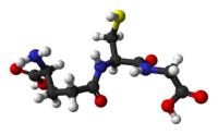
| Glutathione[1] | |
|---|---|
 |
|
 |
|
| IUPAC name |
|
| Other names | γ-L-Glutamyl-L-cysteinylglycine (2S)-2-Amino-5-[[(2R)-1-(carboxymethylamino)-1-oxo- 3-sulfanylpropan-2-yl]amino]-5-oxopentanoic acid |
| Identifiers | |
| Abbreviations | GSH |
| CAS number | 70-18-8 |
| PubChem | |
| MeSH | |
| SMILES |
|
| ChemSpider ID | |
| Properties | |
| Molecular formula | C10H17N3O6S |
| Molar mass | 307.32 g/mol |
| Melting point |
195 °C, 468 K, 383 °F |
| Solubility in water | Miscible |
| Except where noted otherwise, data are given for materials in their standard state (at 25 °C, 100 kPa) Infobox references |
|
Glutathione (GSH) is a tripeptide. It contains an unusual peptide linkage between the amine group of cysteine and the carboxyl group of the glutamate side chain. Glutathione, an antioxidant, protects cells from toxins such as free radicals.[2]
Thiol groups are kept in a reduced state at a concentration of approximately ~5 mM in animal cells. In effect, glutathione reduces any disulfide bond formed within cytoplasmic proteins to cysteines by acting as an electron donor. In the process, glutathione is converted to its oxidized form glutathione disulfide (GSSG). Glutathione is found almost exclusively in its reduced form, since the enzyme that reverts it from its oxidized form, glutathione reductase, is constitutively active and inducible upon oxidative stress. In fact, the ratio of reduced glutathione to oxidized glutathione within cells is often used scientifically as a measure of cellular toxicity[citation needed].
Contents |
[edit] Biosynthesis
Glutathione is not an essential nutrient since it can be synthesized from the amino acids L-cysteine, L-glutamic acid and glycine.
It is synthesized in two adenosine triphosphate-dependent steps:
- First, gamma-glutamylcysteine is synthesized from L-glutamate and cysteine via the enzyme gamma-glutamylcysteine synthetase (a.k.a. glutamate cysteine ligase, GCL). This reaction is the rate-limiting step in glutathione synthesis.
- Second, glycine is added to the C-terminal of gamma-glutamylcysteine via the enzyme glutathione synthetase.
Glutamate cysteine ligase (GCL) is a heterodimeric enzyme composed of a catalytic (GCLC) and modulatory (GCLM) subunit. GCLC constitutes all the enzymatic activity, whereas GCLM increases the catalytic efficiency of GCLC. Mice lacking GCLC (i.e., all de novo GSH synthesis) die before birth.[3] Mice lacking GCLM demonstrate no outward phenotype, but exhibit marked decrease in GSH and increased sensitivity to toxic insults.[4][5][6]
While all cells in the human body are capable of synthesizing glutathione, liver glutathione synthesis has been shown to be essential. Following birth, mice with genetically-induced loss of GCLC (i.e., GSH synthesis) only in the liver die within 1 month of birth.[7]
The biosynthesis pathway for glutathione is found in some bacteria, like cyanobacteria and proteobacteria, but is missing in many other bacteria. Most eukaryotes synthesize glutathione, including humans, but some do not, such as Leguminosae, Entamoeba, and Giardia. The only archaea that make glutathione are halobacteria.[8][9]
[edit] Function
Glutathione exists in reduced (GSH) and oxidized (GSSG) states. In the reduced state, the thiol group of cysteine is able to donate a reducing equivalent (H++ e-) to other unstable molecules, such as reactive oxygen species. In donating an electron, glutathione itself becomes reactive, but readily reacts with another reactive glutathione to form glutathione disulfide (GSSG). Such a reaction is possible due to the relatively high concentration of glutathione in cells (up to 5 mM in the liver). GSH can be regenerated from GSSG by the enzyme glutathione reductase.
In healthy cells and tissue, more than 90% of the total glutathione pool is in the reduced form (GSH) and less than 10% exists in the disulfide form (GSSG). An increased GSSG-to-GSH ratio is considered indicative of oxidative stress.
GSH is known as a substrate in both conjugation reactions and reduction reactions, catalyzed by glutathione S-transferase enzymes in cytosol, microsomes, and mitochondria. However, it is also capable of participating in non-enzymatic conjugation with some chemicals, as in the case of N-acetyl-p-benzoquinone imine (NAPQI), the reactive cytochrome P450-reactive metabolite formed by paracetamol (or acetaminophen as it is known in the US), that becomes toxic when GSH is depleted by an overdose of acetaminophen.
Glutathione conjugates to NAPQI and helps to detoxify it, in this capacity protects cellular protein thiol groups, which would otherwise become covalently modified; when all GSH has been spent, NAPQI begins to react with the cellular proteins, killing the cells in the process. The preferred treatment for an overdose of this painkiller is the administration (usually in atomized form) of N-acetyl-L-cysteine, which is processed by cells to L-cysteine and used in the de novo synthesis of GSH.
Glutathione (GSH) participates in leukotriene synthesis and is a cofactor for the enzyme glutathione peroxidase. It is also important as a hydrophilic molecule that is added to lipophilic toxins and waste in the liver during biotransformation before they can become part of the bile. Glutathione is also needed for the detoxification of methylglyoxal, a toxin produced as a by-product of metabolism.
This detoxification reaction is carried out by the glyoxalase system. Glyoxalase I (EC 4.4.1.5) catalyzes the conversion of methylglyoxal and reduced glutathione to S-D-lactoyl-glutathione. Glyoxalase II (EC 3.1.2.6) catalyzes the hydrolysis of S-D-lactoyl-glutathione to glutathione and D-lactic acid.
Glutathione has recently been used as an inhibitor of melanin in the costmetics industry. In countries like the Phillipines, this product is sold as a whitening soap. Glutathione dose dependently inhibited melanin synthesis in the reaction of tyrosinase and L-DOPA. The inhibition of melanin synthesis was recovered by increasing the concentration of L-DOPA, but not recovered by increasing tyrosinase. Glutathione inhibited the binding between tyrosinase and L-DOPA. Although the synthesized melanin was aggregated within 1 h, the aggregation was inhibited by the addition of glutathione. These results indicate that glutathione inhibits the synthesis and agglutination of melanin by interrupting the function of L-DOPA. "Cite error: Closing </ref> missing for <ref> tag[10]
However, tissue and sperm glutathione concentrations can be raised by increased intake of the precursor cysteine, or in chronic conditions, by S-adenosylmethionine (SAMe).[11][12] Glutathione precursors rich in cysteine include N-acetylcysteine (NAC)[13][14] and undenatured whey protein,[15][16][17][18][19][20][21][22] and these supplements have been shown to increase glutathione content within the cell. N-Acetylcysteine is available both as a drug and as a generic supplement.
There is no evidence that glutathione has any effect in skin whitening.[citation needed]
[edit] Pathology
Excess glutamate at synapses, which may be released in conditions such as traumatic brain injury, can prevent the uptake of cysteine, a necessary building block of glutathione. Without the protection from oxidative injury afforded by glutathione, cells may be damaged or killed. [23]
[edit] See also
- Glutathione synthetase deficiency
- Ophthalmic acid
- roGFP, a tool to measure the cellular glutathione redox potential
[edit] References
- ^ Merck Index, 11th Edition, 4369.
- ^ Pompella A, Visvikis A, Paolicchi A, De Tata V, Casini AF (Oct 2003). "The changing faces of glutathione, a cellular protagonist". Biochem Pharmacol. 66 (8): 1499–503. doi:. PMID 14555227. http://linkinghub.elsevier.com/retrieve/pii/S0006295203005045.
- ^ Dalton, TP; et al. (2000). "Knockout of the Mouse Glutamate Cysteine Ligase Catalytic Subunit (Gclc) Gene: Embryonic Lethal When Homozygous, and Proposed Model for Moderate Glutathione Deficiency When Heterozygous". Biochem Biophys Res Commun. 279 (2): 324. doi:.
- ^ Yang Y, et al. (2002). "Initial Characterization of the Glutamate-Cysteine Ligase Modifier Subunit Gclm(-/-) Knockout Mouse. NOVEL MODEL SYSTEM FOR A SEVERELY COMPROMISED OXIDATIVE STRESS RESPONSE". J Biol Chem 277 (51): 49446. doi:. PMID 12384496.
- ^ Giordano G, et al. (2007). "Organophosphorus insecticides chlorpyrifos and diazinon and oxidative stress in neuronal cells in a genetic model of glutathione deficiency". Toxicol Appl Pharmacol 219 (2-3): 181. doi:.
- ^ McConnachie LA, Mohar I, Hudson FN, et al (Oct 2007). "Glutamate cysteine ligase modifier subunit deficiency and gender as determinants of acetaminophen-induced hepatotoxicity in mice". Toxicol Sci. 99 (2): 628–36. doi:. PMID 17584759.
- ^ Chen Y, et al. (2007). "Hepatocyte-specificGclc deletion leads to rapid onset of steatosis with mitochondrial injury and liver failure". Hepatology 45: 1118. doi:.
- ^ Shelley D. Copley and Jasvinder K. Dhillon (2002). "Lateral gene transfer and parallel evolution in the history of glutathione biosynthesis genes" (free full text). Genome biology 3: research0025.1. doi:. http://genomebiology.com/2002/3/5/RESEARCH/0025.
- ^ Grill D, Tausz T, De Kok LJ (2001). Significance of glutathione in plant adaptation to the environment. Springer. ISBN 1402001789. http://books.google.com/books?hl=sv&lr=&id=aX2eJf1i67IC&oi=fnd&pg=PA13&ots=8feo-QOEPa&sig=XAMjZ0Wan17vmoUKg_FFNRl8g0I#PPP1,M1.
- ^ AIDS Line Update
- ^ Liber CS, Packer L (Nov 2002). "S-Adenosylmethionine: molecular, biological, and clinical aspects—an introduction". Am J Clin Nutr. 76 (5): 1148S–50S. PMID 12418492.
- ^ Liber CS (Nov 2002). "S-Adenosyl-L-methionine: its role in the treatment of liver disorders". Am J Clin Nutr. 76 (5): 1183S–1187S. PMID 12418503.
- ^ Acetylcysteine and glutathione, PubMed
- ^ Gross CL, Innace JK, Hovatter RC, Meier HL, Smith WJ (1993). "Biochemical manipulation of intracellular glutathione levels influences cytotoxicity to isolated human lymphocytes by sulfur mustard". Cell Biol. Toxicol. 9 (3): 259–67. doi:. PMID 8299004.
- ^ Glutathione information for Physicians
- ^ Micke P, Beeh KM, Schlaak JF, Buhl R (Feb 2001). "Oral supplementation with whey proteins increases plasma glutathione levels of HIV-infected patients". Eur. J. Clin. Invest. 31 (2): 171–8. doi:. PMID 11168457.
- ^ Moreno YF, Sgarbieri VC, da Silva MN, Toro AA, Vilela MM (Feb 2006). "Features of whey protein concentrate supplementation in children with rapidly progressive HIV infection". J. Trop. Pediatr. 52 (1): 34–8. doi:. PMID 16014759.
- ^ Grey V, Mohammed SR, Smountas AA, Bahlool R, Lands LC (Dec 2003). "Improved glutathione status in young adult patients with cystic fibrosis supplemented with whey protein". J. Cyst. Fibros. 2 (4): 195–8. doi:. PMID 15463873.
- ^ Micke P, Beeh KM, Buhl R (Feb 2002). "Effects of long-term supplementation with whey proteins on plasma glutathione levels of HIV-infected patients". Eur J Nutr 41 (1): 12–8. doi:. PMID 11990003.
- ^ Bounous G, Baruchel S, Falutz J, Gold P (Jun 1993). "Whey proteins as a food supplement in HIV-seropositive individuals". Clin Invest Med 16 (3): 204–9. PMID 8365048.
- ^ Bounous G, Gold P (Aug 1991). "The biological activity of undenatured dietary whey proteins: role of glutathione". Clin Invest Med 14 (4): 296–309. PMID 1782728.
- ^ Bounous et al. Multiple references on glutathione enhancement with bioactive whey protein in multiple disease states
- ^ Pereira C.F, de Oliveira C.R. (Jul 2000). "Oxidative glutamate toxicity involves mitochondrial dysfunction and perturbation of intracellular Ca2+ homeostasis". Neuroscience Research 37 (3): 227–36. doi:.
[edit] Related research
- Drevet JR (May 2006). "The antioxidant glutathione peroxidase family and spermatozoa: a complex story". Mol Cell Endocrinol. 250 (1-2): 70–9. doi:. PMID 16427183.
- The Role of Glutathione in Cell Defense.
- Wu G, Fang YZ, Yang S, Lupton JR, Turner ND (01 Mar 2004). "Glutathione metabolism and its implications for health". J Nutr. 134 (3): 489–92. PMID 14988435. http://jn.nutrition.org/cgi/pmidlookup?view=long&pmid=14988435.
|
|||||||||||||||||||||||||||||||||||||||||||||||||||||||||||||||
|
||||||||||||||

